In vivo miniscopes

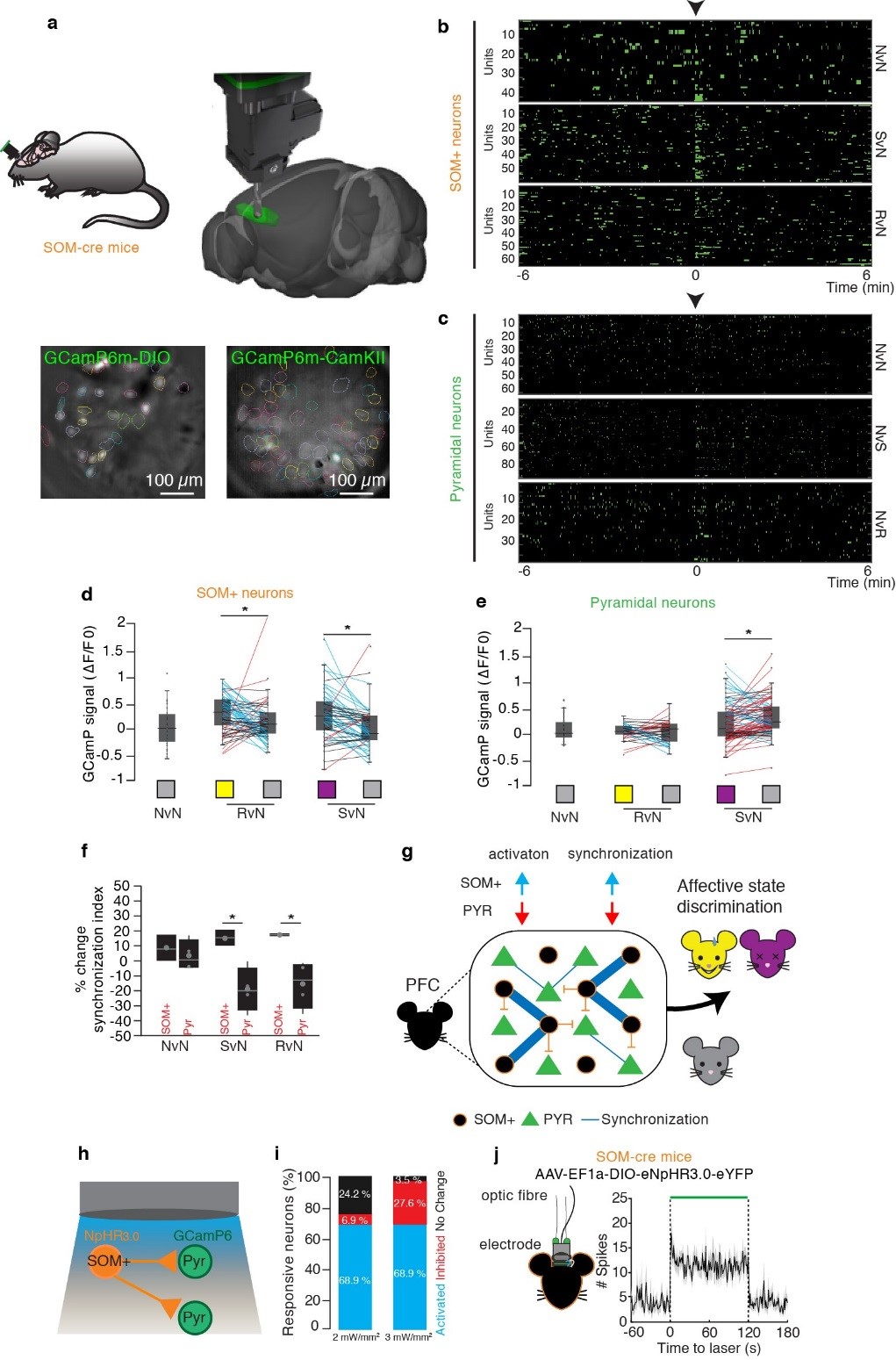
Concurrent activation of SOM+ interneurons in the mPFC is associated with emotion discrimination. (a) SOM:cre mice were injected in the mPFC with AAV.Flex.GCaMP6m to target somatostatin interneurons or injected in the mPFC with both AAV-CamKII-GCamP6f and AAV-DIO-eNpHR3.0 to target pyramidal neurons. Mice were implanted with a ProView lens to allow large scale neuronal Ca2+ imaging using a miniature fluorescence microscope. Cluster traces of Ca2+ transients events from (b) SOM+ and (c) pyramidal neurons during habituation and EDT testing (the black arrow represent the beginning of the test) in three different conditions: with two neutral demonstrators (NvN), a relieved and a neutral (RvN), and a stressed and a neutral (SvN) demonstrators. (d) Normalized Ca2+ signal from SOM+ interneurons during each contact with each demonstrator show an increased activity when exploring an emotionally-altered conspecific compared to a neutral both in the relief and stress conditions. (e) Normalized Ca2+ signal from pyramidal neurons during contact with each demonstrator show no changes between the neutral and relief, while a decrease between a neutral and stress was observed. (f) % of correlation index change between the test and the habituation: analysis of the correlation index (CI) for all possible pairs revealed a significant decrease for each pyramidal pairs in presence of an emotionally altered mice while the CI increases in SOM+ following the interaction with an emotionally altered mouse. (g) Graphical model scheme of the suggested mechanism implicating mPFC SOM+ interneurons in emotion discrimination. Our data converge indicating that the expression of emotion discrimination is driven by a synchronized activation of SOM+ neurons in the mPFC which results in a direct inhibition of a big majority of mPFC pyramidal neurons. Indeed, blocking the SOM+ synchronized activation completely abolished emotion discrimination. Conversely, artificially inducing a SOM+ synchronized activation by optogenetics was sufficient to recapitulate an emotion discrimination. In contrast, a reduced synchronized activation of pyramidal neurons is associated with emotion discrimination, and a direct simultaneous inhibition of mPFC pyramidal neurons by optogenetics is not sufficient to completely abolish emotion discrimination. In agreement, the mPFC SOM+ photostimulation inducing social discrimination inhibit pyramidal neurons, while the mPFC SOM+ photoinhibition abolishing emotion discrimination mostly activated pyramidal neurons. (h) SOM-cre mice injected in the mPFC with AAV-CaMKIIa-GCaMP6f and AAV-DIO-eNpHR3.0 to record calcium dynamics of pyramidal neurons following inhibition of SOM+ neurons. (i) Diagram illustrating the state of mPFC pyramidal neurons during photoinhibition of SOM+ interneurons in the mPFC. During photoinhibition of SOM+ interneurons with either 2 or 3mW/mm2, the same pyramidal neurons (68.9%) shows an increase of their event rate, while variable small minority of pyramidal neurons were inhibited or unaffected. (j) Optogenetic-assisted tetrode recordings in the mPFC in AAV-EF1a-DIO-eNpHR3.0-eYFP injected SOM-cre mice; example of a peristimulus time histogram showing the firing rate (mean ± SEM), of a slow-firing putative pyramidal cells, before, during and after the delivery of the green light pulse (120 s). Modified from Scheggia, Managò et al, Nature Neuroscience, In Press